During the last decades, many breakthroughs in cancer diagnostics and treatment came from new discoveries in basic science. However, despite enormous progress, not all expectations have been fulfilled. Even 50 years after the declaration of the "war on cancer" by the Nixon administration and 20 years after the completion of the human genome, we are far from a comprehensive understanding of the biology of cancer. This is reflected in a shockingly high failure rate of new cancer drug candidates in clinical trials of more than 95%.
The importance of supracellular organization and spatial context of molecular phenomena
In our view, one of the explanations for this failure rate is that many new drug candidates are developed based on mechanistic hypotheses developed outside the organismic or tissue context of the tumor. Therefore, the influence of immune cells, tumor stroma and many other aspects of human physiology cannot be effectively predicted before the clinical trial stage, and furthermore, many potentially targetable cancer properties that emerge at the supracellular level are completely neglected. To improve this situation and more effectively utilize the explosion in knowledge coming from recent technological innovations (in sequencing, proteomics, single-cell technologies), we need to interpret the enormous amounts of data that these techniques generate in their original tissue context. In the last few years, this has become more and more evident in the scientific community and is reflected in many new technologies like spatially resolved transcriptomics (Method of the Year 2020, Nature Methods) and spatial transcriptomics, as well as severalrevolutionarytissueimagingtechnologies developed in the last decade.
Pathology as the "science of spatial context"
Spatial context of cells in tissues has always been the main area of study and expertise of pathologists. In fact, the centuries-old technology of microscopic observation of H&E stained tissue sections can be considered the first spatial technology with single cell resolution. The fact that this simple method is still the most important and by far most cost-effective tool in cancer diagnostics today is an impressive testament to the effectiveness of the approach to evaluate tumor phenotype within its tissue context.
The new role of pathology as a central discipline in spatial "omics"
We believe that the recent realization across the translational research community that spatial context is essential to understand tumor behavior will strengthen the role of pathology as an essential discipline at the interface between basic science and clinical disciplines. We combine cutting-edge methods from basic science with profound expertise in traditional surgical pathology to better understand the biology of cancer, develop better predictive biomarkers and help to improve cancer therapy.
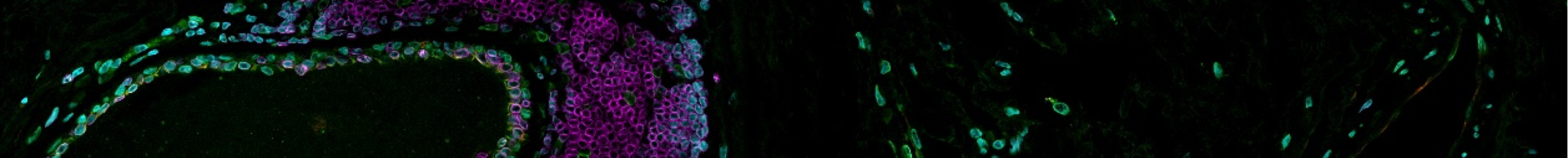
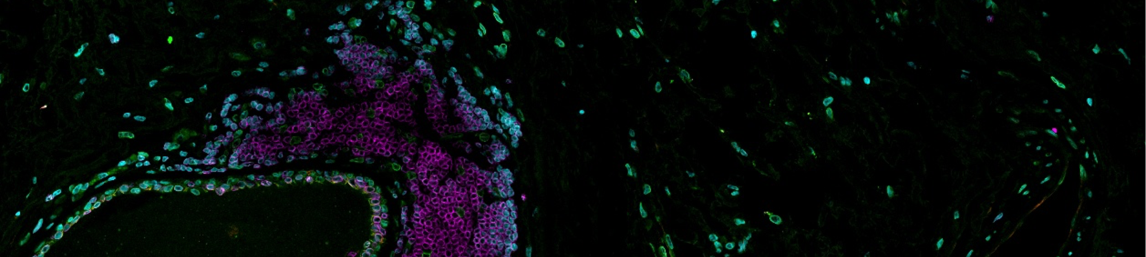
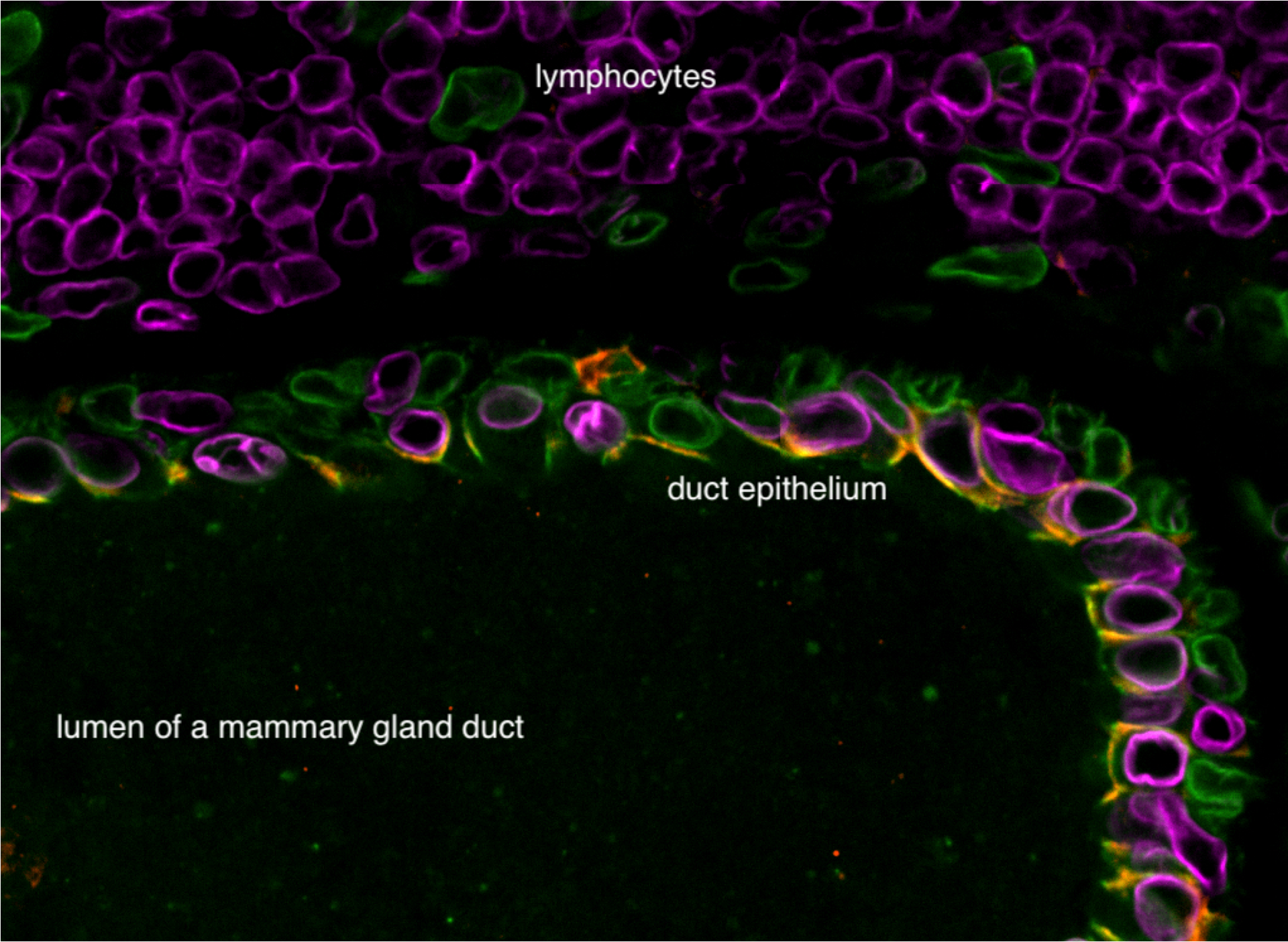
An example of the complex supracellular organization of healthy tissue - section of a normal mammary gland duct with typical bilayered epithelium consisting of luminal cells and basal myoepithelial cells. The surrounding connective tissue contains an aggregation of lymphocytes forming a lymphoid follicle.
Identification of therapeutic biomarkers through characterization of tumor cell heterogeneity in breast cancer and lung cancer at the single-cell-level
Even if they are highly effective or potentially curative, most cancer therapies only work in a subset of patients. Prognostic and therapeutic biomarkers help us to select the most effective therapy option for cancer patients and thus make sure that patients do not have to suffer from unnecessary drug toxicity. For some cancer drugs, e.g. kinase inhibitors that target mutated proteins, we have very good - and obvious - therapeutic biomarkers, like the Bcr-Abl fusion for Glivec. For other cancer drugs, especially the ones which have more complex mechanisms of action, like immune checkpoint inhibitors or chemotherapy drugs, classic monoparametric biomarkers fail to accurately predict therapeutic outcome. New -omics methods allow characterization of tumor tissues at multiple levels with unprecented depth. In part through collaborations, in part in our own lab, we use proteomics, transcriptomics and genomics in combination with multiplexed immunofluorescence imaging in order to develop multilevel spatial therapeutic and prognostic cancer biomarkers. Interpretation of these multidimensional datasets is facilitated through deep learning-based approaches. We aim to recapitulate our findings in realistic ex vivo experimental models such as organoids and use such experiments to better understand cancer cell behavior.
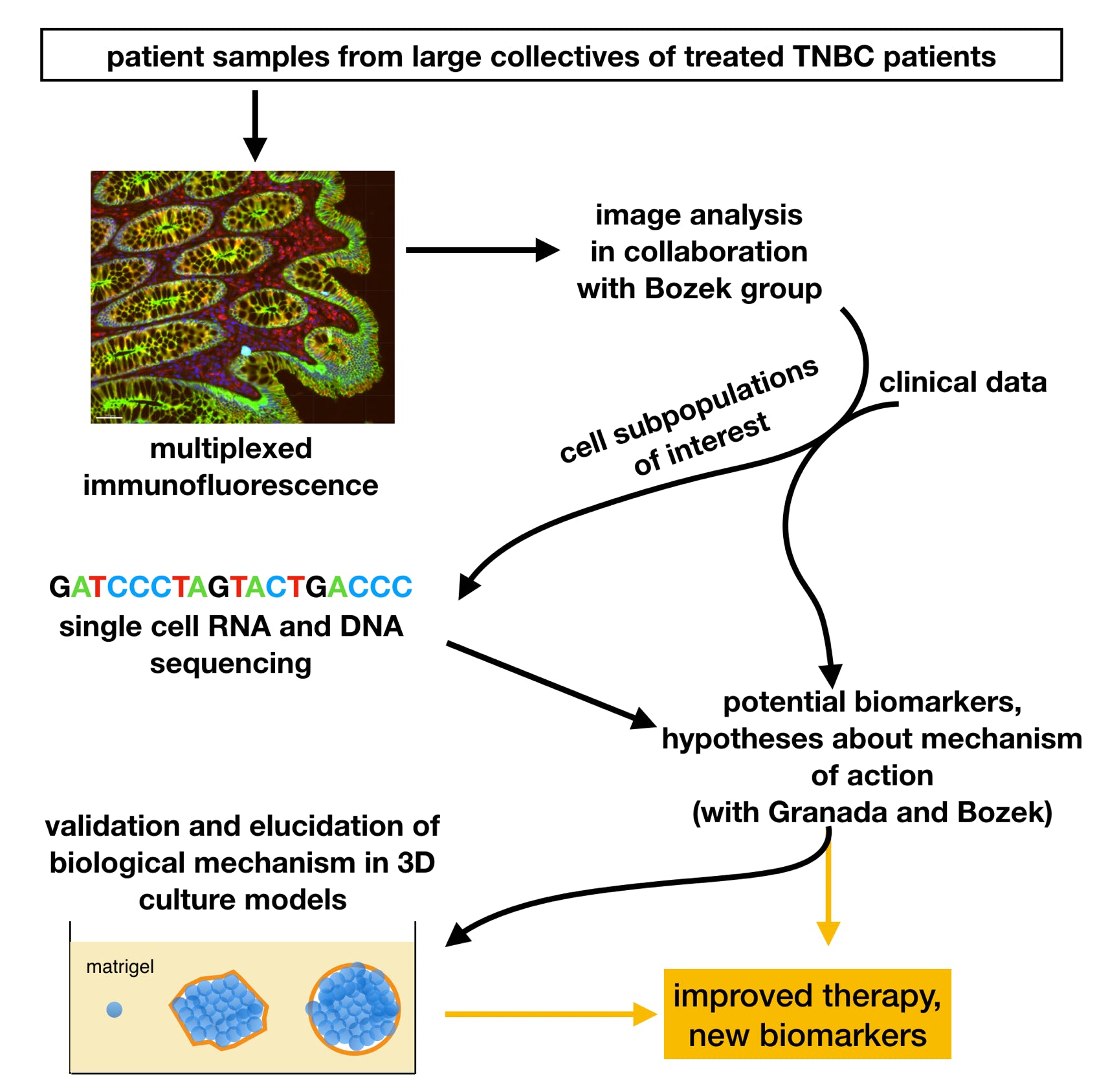
Overview of our project within the DeepLTNBC Junior research network focused on developing new complex biomarkers to better predict the outcome of therapy in triple negative breast cancer (TNBC).
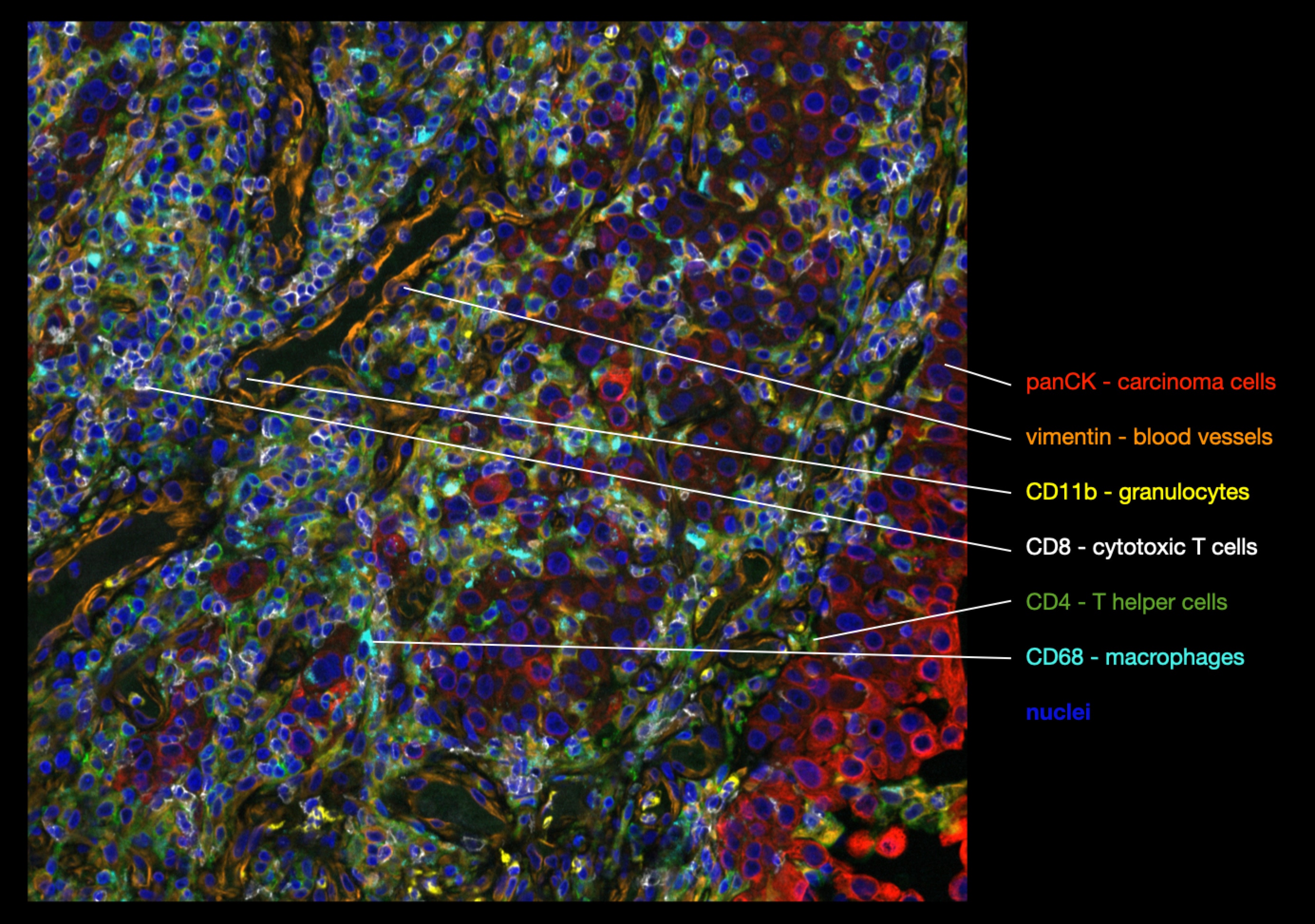
A lung tumor invading the surrounding stroma. Here, we performed multiplexed immunofluorescence to characterize the composition of the tumor-surrounding immune infiltrate. Note the yellow granulocytes transmigrating through the blood vessel walls (orange) into the surrounding stroma.
Microtubule binding drugs and role of microtubule organization in cancer
Microtubule binding drugs like Paclitaxel and Vinca alkaloids are among the most important chemotherapy drugs and cancer drugs in general. They are used to treat a wide range of solid tumors and hematologic malignancies. Since they were discovered empirically, despite their clinical importance, their mechanism of action in patients remains unknown, with several competing hypotheses. This prevents rational development of new drugs using the same mechanism of action. It also makes it difficult to predict which patients will benefit from MT binding drugs, leading to unnecessary drug toxicity and patient suffering as well as a waste of resources in the healthcare system.
Originally, it was hypothesized that this drug class arrests and kills cells during cell division. However, new targeted drugs that were designed to recapitulate this effect lacked the clinical efficacy of MT binding drugs. This raises the question how this so important drug class kills tumor cells in patients.
We are using live cell imaging of mammary organoids combined with high-throughput characterization of breast tumors using multiplexed immunofluorescence, deep-learning based image analysis and third generation sequencing to learn how the conformation and organization of microtubules in tumor cells, as well as their dynamic properties, affect response to this important drug class.
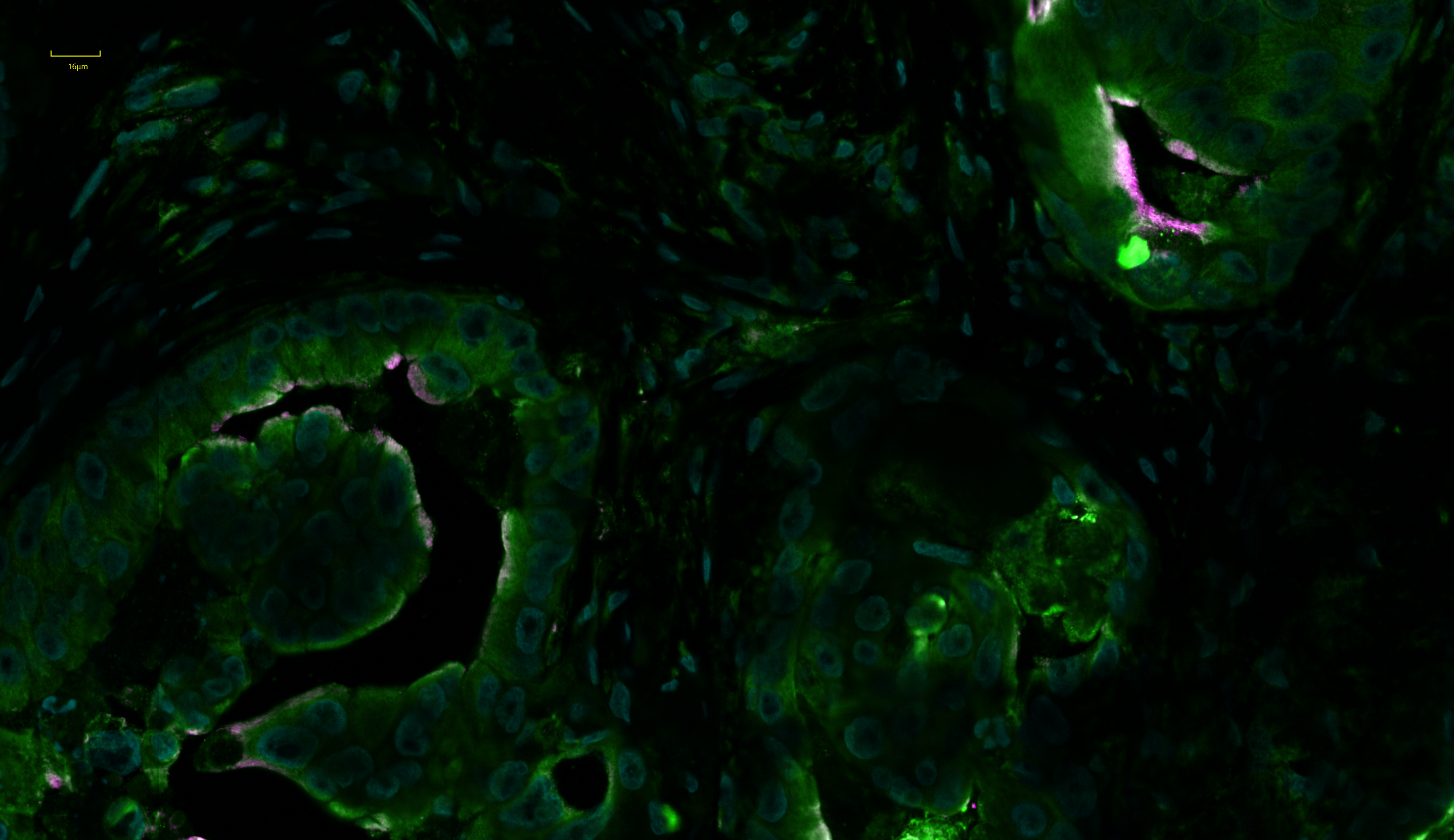
Staining of a triple negative, well-differentiated breast cancer with antibodies against posttranslationally modified tubulin.